I’m back! I brought
movies! Be excited.
So, I’m
currently working on a paper that refuses to die. In the past few weeks, I’ve had to revisit
some old techniques to generate more data (that will hopefully statistically say
what I want – fingers crossed!). While
doing so, I’ve generated some very pretty images of cells that I’d like to
share with you.
In my “You use that inlab?” post, I discussed a little bit about how we grew cells on coverslips, put it on
a microscope slide (painted it with nail polish!) and then looked at our cells under a
microscope. In Figure
63.2, I showed a side view of the cells.
Many people think of cells as flat, two dimensional beings because that
is how they are always drawn in pictures.
Obviously, cells exist in a three dimensional world and have, in
addition to length and width, height.
When these
slides that we create are placed on a standard microscope and we look through
the eyepiece, we are seeing light coming from all heights in the cell. From the tippy top of the cell all the way
through the cellular structures, we are seeing all these things inside a cell
piled on top of each other. Imagine flying
over a high-rise building made of glass.
Looking with a regular microscope is the same as simply looking down
from your helicopter and seeing the building: you’ll see the top and some
floors beneath, but it will be hard to discern what is on different floors with
any clarity.
Wouldn’t
it be cool if we had a microscope that allowed us to look at a very thin slice
of a cell at any height? Pretend you
could cut the cell into ten pieces with a teeny little knife and then look at
each individual slice. It would be the
same as seeing each individual floor within the glass building without having to
look through the other floors. Wouldn’t that be interesting?
It turns out such a microscope
exists! It’s called a confocal microscope and this is the instrument I
have spent nearly eight hours on in the past week. The thing is so smart that I feel like an
idiot using it and am often nervous that I’m breaking it. Today, I was so overwhelmed with all the
settings I had to change from the previous user that I simply restarted the
program. In truth, what I’m doing with
the confocal is elementary; other users in my lab take live images of fruit fly
ovaries depositing eggs. I have no idea
how to do this, but I can look at cells I’ve grown in a petri dish!
I’m
going to show you some of those images now, but first I want to re-acquaint you
with the major parts of a cell and introduce you to an interesting cell organelle called the Golgi body (always with a capital
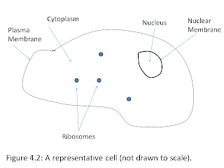
G). Figure
4.2 (woo
– way back!) shows you some of the major cell parts. This picture is representing a cell at one
particular height. Remember that the nucleus,
ribosomes, etc. all have height in addition in length and width. Suffice it to say that what is shown is not
exhaustive of all the cell parts. Of the
many things left out, one is the Golgi body, which I did go back and draw in
for you in Figure 70.1. It’s a big, amorphous looking thing, isn’t
it?
The
Golgi body (also
called the Golgi complex or the Golgi apparatus) is responsible for
packaging proteins that need to be sent outside the cell’s plasma
membrane. It’s the shipping
department. How it does this is not important,
but it’s a necessary and important organelle within the cell.
The
cells I looked at in the confocal this week have their Golgi bodies marked with
a fluorescent protein. Remember
fluorescent proteins – I talked about them in my Fluorescent Proteins post? If not, the summary is that the Golgi body in
these cells will glow in a blu-ish color* due to the presence of cyan
fluorescent protein there.
With
confocal microscopy allowing us to look at a cell at all heights and a fluorescent
protein highlighting the Golgi body for us, we can fly through a cell. Video 70.1 takes
you from the top of a cell all the way through it to the bottom of the
cell. There is one cell in the middle of the screen. The Golgi will be quite blue and the plasma membrane is only poorly outlined, but you should be able to clearly see a cell nonetheless. I’ve tried to schematically
explain this in Figure 70.2. I could have marked many different things in
a cell; it did not have to be the Golgi body.
I could have chosen the nucleus, the plasma membrane, small membrane
bodies called endosomes, or even large protein complexes. The choices of colors and ways to mark a
protein are somewhat endless – this is just what my project demanded.
Enjoy
the ride!
Confocal miscroscope – a specialized
microscope that allows for seeing “slices” of a cell. These slices can then be stacked together to
make a video of a three-dimensional cell (also called a z-stack).
Organelle – Specialized areas
in cells that have specific functions.
Example: nucleus, Golgi Body, ribosomes, vesicles, endoplasmic
reticulum, etc
* - The camera on the confocal only detects photons, not
color – I’ve added in the color.
REFERENCES
Alberts et al. “Molecular Biology of the Cell, 4th
Edition.” Garland Science, New York, New
York. (2002).


